The Contrast Sensitivity Function (CSF) and Image Discrimination
Eli Peli
Schepens Eye Research Institute, Harvard Medical School, Boston, MA 02114
and
New England Eye Center, Tufts University School of Medicine, Boston, MA 02111
Abstract
In a previous study the simulation of image appearance from different distances was shown to be effective
1 . The simulated observation distance accurately predicted the distance at which the simulated image could be discriminated from the original image. Due to the 1/f nature of natural images spatial spectra, the individual CSF used was actually tested only at one retinal spatial frequency. To test the CSF relevant for the discrimination task over a wide range of frequencies, the same simulations and testing procedure were applied to 5 contrast versions of the images. The lower contrast images probe the CSF at lower spatial frequencies, while higher contrast images test the CSF value at higher spatial frequencies. Images where individually processed for each of 4 observers using their individual CSF to represent the appearance of the images from 3 distances where they span 1, 2, and 4 deg of visual angle, respectively. Each of the 4 pictures at the 5 contrast levels and the 3 simulated distances was presented 10 times side-by-side with the corresponding original image. Images were observed from 9 different observation distances. Subject task was to determine which of the two was the original, unprocessed image. For each simulated distance the data was use to determine the discrimination distance threshold.
Results of testing using simulations calculated with CSFs measured from a 2 m where veridical for the images in the 30 - 100% contrast range. The 10% image was discriminated at distances larger than the simulated distances. The 300% image was discriminated at a shorter distance. A second set CSFs were obtained from a range of observation distances (0.5 m to 8 m), to overcome limitations of the display. These data showed higher sensitivity for low frequencies and lower sensitivity at the higher frequencies as predicted by the simulation testing results. Replication of the simulation experiments with the combined CSFs resulted in a much better prediction of the discrimination distances. Thus, the CSF relevant for the image discrimination task was verified over a wide range of spatial frequencies while further validating the visual model and its use for simulations.
1. Introduction
Simulating the appearance of a scene or an image to an observer is a useful design and analysis tool. Such pictorial representations have been attempted by many investigators over the years in a variety of applications in vision science2,3,4,5 and engineering6,7,8. Such simulations are frequently generated within the context of a computational vision model. One such multi-scale model of spatial vision was used to calculate local band-limited contrast in complex images9. This contrast measure, together with observers' contrast sensitivity functions (CSFs), expressed as thresholds, was used to simulate the appearance of images to observers, taking into account many of the non-linearities inherent in the visual system. Using the CSFs of low vision patients, the simulation was applied to the design of image enhancement algorithms for the visually impaired8. The local band limited contrast model was also used to simulate the appearance of images presented to the peripheral retina10 using the CSF measured at various retinal eccentricities. Others have applied the same concept of local band-limited contrast with small variations7,11,12 and found it useful in comparing image quality12 and in other applications7 .
In a recent study Peli1 tested and demonstrated the validity of the central (foveal) visual model using simulations of the appearance of complex images generated with the model. Images generated with the model represented the appearance of images from various observation distances. Observers view the images simulated using their individual CSF from a wide range of distances side-by-side with the original image and attempted to discriminate the original from the simulated image. The distance at which discrimination performance was at threshold was compared with the simulated observation distance. That study also sought to determine what CSF data should be used in this or any other vision models of this type. While methodological changes can account for the variability of CSF data in the literature13, we do not yet know which of these is the appropriate method for determining CSFs that represent complex image perception in the context of pyramidal multi-scale vision models. That study demonstrated that the CSF obtained with a fixed aperture of grating could be rejected when compared with CSF obtained with fixed bandwidth Gabor patches stimuli. Further, it showed that CSF obtained with orientation discrimination task could be rejected while the CSF obtained with a detection task could not be rejected. In both cases 1-octave Gabor patches were used as stimuli. The variable distance simulation and testing method were also shown to be sensitive, affected by small differences as induced by the high frequency residual. The main limitation of the previous study was the fact that with the methodology used the validity of the CSF was tested only at one spatial frequency, as explained below. This paper sought to expand this investigation over a wide range of retinal spatial frequencies.
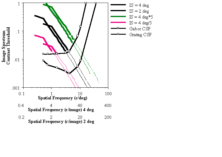
In applying the vision model to simulations or other applications one needs to consider both the object's contrast spectrum computed in terms of cycles/object or cycles/image and an observer's CSF expressed in terms of cycles/deg. To derive the object's spectrum at the retina (Fig. 1, solid black curve) the distance of the object to the observer needs to be known. Any information in the image that falls below the observer's threshold (i.e., below the point at which the contrast threshold curve intersects the image spectrum curve) is treated by the model as not visible to the observer. To illustrate this, the simulation should remove all that information. This is illustrated by the spectrum line turning to a thin line at the values that are below threshold. If the original and simulated images are viewed from the simulated distance or farther, they should be indistinguishable because the same information from the original that would be lacking due to the visual response was removed in the simulation as well. However, if the original and simulation are viewed from a closer distance, the difference in content between the original and the simulation should be visible.
As the size of displayed object shrinks when the distance of the object from the observer increases, its retinal spatial frequencies increase. It was previously thought by us14 and others15 that this change results in a shift of the spectrum to the right along the spatial frequency axis (in Fig.1). However, as Brady and Field16 pointed out, the spectrum actually shifts both to the right (higher frequencies) and down (lower contrast) sliding along the line with a slope of -1.0. Most natural images have a spatial frequency spectrum that behaves as 1/f, which also have a slope of -1.0. Thus a change in object size causes the spectrum to "slide along itself" (Fig. 1, dashed black curve). As a result, the spectrum of the farther image intersects the CSF curve at a essentially the same retinal frequencies. Only the mapping of the relevant object frequencies to retinal frequencies changes. Therefore, the experiments in Peli1 have probed only a very limited range of spatial frequencies in the CSF. To examine the CSF at other frequencies one needs to use images whose spectra intersect the CSF at other frequencies. This was achieved in the current study by using higher and lower contrast versions of the same images as illustrated schematically in Fig 1. In fact it was the amplitude of the images that was increased or decreased not the contrast. This operation in which the image dc value is subtracted and the remaining values are scaled up or down is frequently referred to as contrast increase or decrease. As pointed by Peli9 the changes in contrast are equivalent to changes in amplitude only where the local luminance is equal to the mean luminance. I will use the terms contrast changes here in conforming to previous use and in recognition that in many places the differences are small. The lower contrast images' spectra intersect the threshold CSF curve at lower spatial frequency and thus can be used to test the CSF at lower frequencies. Similarly the higher contrast image was used to test the CSF at a higher spatial frequency.
|
|
Fig. 1. The interaction of image spatial frequency content (at different image contrasts (amplitudes)) with the observer's CSF from 2 different observation distances. Solid black curve represents a typical image spectrum (1/f) for an image spanning 4 deg. The part of the spectrum below the observer's CSF (detection threshold obtained with Gabor stimuli) will not be detectable. A change in observation distance which causes the image to shrink to 2 deg. shifts the spectrum along the slope of -1/f (dashed black line). At the new distance lower object frequencies are removed by the observer's CSF but essentially the same retinal frequencies are involved. The gray curves represent the spatial spectra of images with increased and decreased contrast which shift the intersection of the spectra with the threshold to higher and lower retinal frequencies, respectively, enabling testing of other parts of the CSF.
Figure 1 is useful only to illustrate the logic of the experiments described below. The analysis it represents cannot replace the information we seek from the simulations and from direct testing of the simulation. The effects of contrast threshold on apparent contrast in the images are local, not global. Thus the effective contrast is not accurately represented by the (1-D) radially averaged amplitude spectrum, because in the simulations we were working with local contrast, not amplitude9, and the simulation algorithm is not represented accurately by the linear filtering depicted in the schematic.
2. Methods
The simulations were tested by presenting the original image side-by-side with the simulation of its appearance from a certain distance. If the simulations are valid, the simulated image and the original should be indistinguishable from a distance equal to or farther than the distance assumed in the simulation14. The two images should be progressively easier to distinguish at distances shorter than the simulated distance.
Observers viewed image pairs from various distances and were asked to make a forced-choice distinction between the simulated and the original image. The simulated images used to test each observer were calculated using her/his individual CSF. Four different images at 5 different contrast versions were used in this experiment. For each image, three simulated views were generated representing views from three different distances. For the three simulated observation distances (106, 212, and 424 cm), the images spanned visual angles of 4, 2, and 1 deg respectively. The simulated distance and the corresponding span in degrees serves to establish the proper relations between the subject's CSF expressed in c/deg and the image spatial content expressed in terms of c/image. The subjects viewed the image pairs from nine distances, including shorter (53 cm) than the shortest simulated distance and longer (848cm) than the longest simulated distance. Each image at each simulation distance was presented 10 times at each viewing distance. The position of the simulated image relative to the original (right or left) was randomly selected for each presentation. From each observation distance the percent correct identification of the processed/simulated image was calculated for each simulated distance for the 4 images. The data for each simulated distance (percent correct out of 40 responses for each observation distance) was fit with a Weibull psychometric function to determine threshold at a 75% correct level. The distance at which the subject performed at 75% level was compared to the simulated distance. If the simulations and the CSF used in the simulation represent the subject perception correctly, the measured and simulated distance should be equal.
The CSF data used in the simulations were obtained for each subject individually. The CSFs data were obtained using 1-octave Gabor patches and a simple detection task. The CSF data were collected on a Vision Works system (Durham, NH) using a M21LV-65MAX monitor with DP104 phosphor operating at 117 Hz, non-interlaced. CSF data were collected with both method-of-adjustment (MOA) and staircase procedure, as indicated. The staircase included 2 practice reversals and the threshold was calculated as the average of 4 test reversals. Seven interwoven frequencies separated by 1 octave between 0.5 and 32 c/deg were presented in each block. For the MOA six responses at each frequency were averaged. The stimuli were the same Gabor patches of 1 -octave bandwidth in all cases (vertical orientation only).
The image pairs were presented on a 19 in (48 cm), non-interlaced monochrome video monitor of the Sparc 10 Workstation (Sun Microsystems, Mountain View, CA). Linearity of the display response was obtained using an 8-bit lookup table. The calibrated screen provided a linear response over 2 log units. The images were 256256 pixels each and were presented at the middle of the screen, separated by 128 pixels. The background luminance around the images was set to the mean luminance level of the display (40 cd/m2). The four images used are common images used in image processing. These images were originally recorded with standard video cameras designed to display on a non-linearized CRT. To enable a linear relationship between the displayed luminance levels and the numerical representation of the images, we presented the images using a linearizing (Gamma corrected) lookup table. To maintain the natural appearance and contrast range of the images the original images were preprocessed to include the measured display Gamma function17. The original images were also produced at varying contrasts. This was achieved by subtracting the mean luminance level from the image multiplying each pixel by the corresponding contrast (10%, 30%, 50%, and 300%) and adding the mean luminance back. The 300% contrast image was saturated wherever the dark or bright values exceeded the dynamic range of the display. The simulations for each of the 4 images 5 contrast versions and 3 simulated observation distances were generated as described in Peli1.
Observers were seated in a dimly lit room and adapted to the mean luminance of the display for 5 min. before beginning the experiment. Location of the simulated image (right or Left) was indicated using the right and Left buttons on a mouse. A new pair of images emerged 0.1 sec. after each response and remained on until the subject responded. The order of observation distances was randomized
3. Results
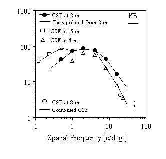
I. Testing the simulations of variable contrast image versions A. Measurements of the CSFThe first experiment was conducted with simulations calculated using CSF data measured from a fixed, 2 m, observation distance. The display size and resolution limited the range of frequencies measured from this distance to 0.5 - 16 c/deg. The CSF values needed for the simulations at frequencies outside this range were extrapolated by extending the low and high frequencies limbs of the CSF linearly14. The CSF was measured using the method-of-adjustment (MOA). For the subjects who were well trained psychophysics subjects, the results with MOA differed only slightly from the CSF obtained under the same condition using the staircase procedure. The CSF data and the standard error of the measurements were similar to data collected for these stimuli with different systems and using forced choice procedures13. This was not the case for one novice subject. For this subject (JML) the staircase procedure data was similar to the other observers, but the MOA data showed substantially reduced sensitivity (as much as 0.5 log units at middle and low frequencies), even when measured repeatedly.
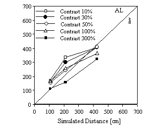
B. Image discriminationsFour observers participated in this experiment and their results were similar. If the simulations were veridical, the fitted curves should have crossed the 75% correct level at the simulated distance and thus all points in Fig. 2 should lay on the diagonal line.. As can be seen the results of the simulation testing were veridical only for the images in the 30- 100% contrast range even for the most practiced subject (AL, who participated in a pervious study1 using the same task). For these moderate contrast images the distance at which the original was distinguished from the simulation was very close to the simulated distance. The 10% image was discriminated at distances larger than the simulated distances, indicating that the CSF values used in the simulations at low frequencies were too low. Stated otherwise, the thresholds implemented in the simulations were too high, removing more of the image than appropriate and thus making the discrimination task easier. The 300% image was discriminated at a shorter distance, indicating that the CSF values used at the high frequencies were too high. The result for a second subject (KB) who was well trained in psychophysical tests, but novice to this task, are shown in Fig 2b. These results are similar in nature except that performance was poorer requiring shorter observation distances to distinguish the simulation image from the originals. In addition, the results for the various contrast versions for this subject differ even more for the moderate contrast versions as compared with the results for AL. The results for two more subjects (not shown) were similar to subject AL's in that they were centered around the diagonal prediction line, although their variability was larger, i.e. of the same order as Subject KB's results.
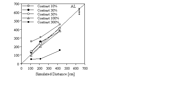
Figure 2. The distances at which the
simulated images were distinguished from the corresponding original images compared with the
simulated observation distance. a) For a well practiced subject the data is deviating from the
prediction only for the extreme contrast conditions corresponding to detection of low spatial
frequencies (10% contrast) and high spatial frequencies (300% contrast). b) For a novice subject
the simulated images were distinguished from the original image at a shorter distance than
simulated. These results illustrate again that, using
this methodology, one can accept or reject the values of the CSF data used for the simulation.
The addition of the image contrast variable in this experiment enable us to test the CSF along a
wider range of frequencies than that tested by Peli1 and note that the sensitivities
at both the low and high end of the range of frequencies were not representative of the observers
perception in the task. In particular the data suggest that individual CSF measured and used in
the simulation under-estimated the observer sensitivity at low spatial frequencies and
over-estimated the sensitivity at high spatial frequencies. Since extrapolated CSF values were
used at both ends of the frequency range in the simulation, further experiments were carried out
to determine if the deviation at low and high contrast was a result of an error introduced
through the extrapolation or the measurements of the CSF.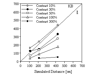
II. Accounting for the deviations from predictions
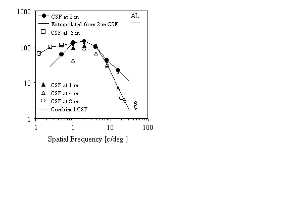
The contrast sensitivity was re-measured for 2 of the 4 subjects using the same system, stimuli, and procedure but the observation distance was varied to enable extension of the tested frequency range. The shortest distance of 0.5 m transferred the lowest frequency tested from 0.5 c/deg to 0.125 c/deg. The 3 lowest frequencies were measured from this distance. The farthest distances of 4 and 8 m permitted testing at frequencies as high as 24 c/deg (our observers could not detect the 32 c/deg stimuli at any contrast). As can be seen in Fig. 3 the results for the low frequencies taken at the shorter distance resulted in higher manifest sensitivity, as was predicted by the simulation results of Experiment 1. The CSF at the high frequencies taken from a longer distance of 4 and 8 m were almost overlapping. These results, at high frequencies, were substantially lower in sensitivity when compared to the data measured and extrapolated from the 2 m measurements. These changes in sensitivity are also consistent with the results obtained in the simulations, suggesting that the contrast sensitivity of the observers in the task is better represented by the CSF values measured (at the corresponding distances) and not those extrapolated from the CSF obtained at 2 m. It should be noted that except for the 24 c/deg conditions the new measurements in all other cases used the same physical stimuli used at the 2 m distance. Possible reasons for the different results are presented in the discussion section.
|
|
|
Figure 3. The contrast sensitivity data measured for 2 subjects at different observation distances. The data collected at 2 m distance together with the illustrated extrapolations were used in Experiment 1. The data shown with a solid line marked "combined CSF" was used in the simulations of Experiment 2.
To verify the simulations effect and the CSF used in the simulation the procedure of Experiment 1 was repeated for two subjects using the CSF obtained by combining the data from the various experiments using the short distance observations for the low spatial frequencies, the 2 m measurements for intermediate frequencies, and the 4 m measurements for the high frequencies. The CSF at 32 c/deg used in the simulations was extrapolated from values at 8, 16, and 24 c/deg, since our observers could not perform the task at that frequency. The simulations were computed using the combined CSF functions presented in Fig. 3 and the testing was repeated. The results, Fig. 4, clearly show a convergence of the data towards the diagonal line for subject AL. Subject KB shows a substantial convergence of the data from various contrast versions and in addition a combined improvement in overall performance. This improvement may be accounted for by the increased familiarity with the task. For both subjects the deviations from the predicted distance of distinguishing the original from the simulation is reduced when compared to the data of Fig. 2. In particular the values for the 10% and 300% contrast images converge towards the other values. The results for the 300% contrast image remain separated from the rest of the samples. Since the 300% images test the CSF at high spatial frequencies, this indicates that the observers' perception in the task is represented by even lower sensitivity than that measured from the 4 m observation distance. The whole set of simulation testing was replicated for one subject (AL) following re-calibration of the display at a brighter mean luminance level. The results were similar to those presented in Fig. 4 except for a small improvement in overall performance.
|
|
|
Figure 4. The distances at which the simulated images were distinguished from the corresponding original images compared with the simulated observation distance, for the same subjects as in Fig 2. Here the simulations were computed using the combined CSFs obtained from different observation distances. a) For the well practiced subject the data with the combined CSF is now very close to the prediction represented with the diagonal solid line. b) For the novice subject the practiced gained in the task results in the simulated images here being distinguished from the original image at a farther distance than simulated. In addition, the different contrast versions are detected closer to each other and closer to the prediction line than in Fig 2b.
4. Discussion
The results of these experiments re-verified that the model proposed by Peli9 and used to simulate the appearance of an image from different observation distances is valid19. The simulated images are distinguishable from the original at distances close to the simulated distances. The size of the effects, as occurs when observer's distance from the display is doubled, is of the magnitude of interest in image quality metrics. Since we are able to accurately simulate such effects using the vision model employed here it stands to reason that such models could be successfully employed to calculate such differences in order to estimate image quality7,12. In addition, the current study has demonstrated that this method can be used to test the applicability of a specific empirically derived CSF to the performance of a discrimination task with complex images.
Of particular interest in this work is the possible reasons for the differences between the CSF's obtained at different frequencies. The low frequency end is simple to account for. The low frequency Gabor patches used from a distance of 2 m are quite large physically occupying substantial part of the screen. The edge of the screen (outside the active video range) is dark and creates a high contrast feature which when close to the patch may reduce its visibility. Moving the observer closer to the screen reduces the physical size of the patches on the screen for the same spatial frequencies and thus increases their distance from the edge and reduces its masking effect. Indeed for both subjects the detection threshold for the three lowest spatial frequencies was almost equal at 2 m and 0.5 m (which were the same physical stimuli) suggesting that the reduction in sensitivity for these Gabor patches at low frequency is only a masking effect. This argue for even higher sensitivity at low frequency than represented by the "combined CSF" in Fig. 3.
The explanation for the change in CSF for high spatial frequencies we found with increase in distance is not as obvious. Although the high spatial frequency targets are physically small on the screen at 2 m distance there is sufficient resolution to adequately represent the Gabor patch (about 8 pixel/cycle). Even if this resolution is too low it is not clear why it would lead to higher apparent sensitivity. In fact the opposite effect may be expected. Using the larger stimuli from 4 or 8 m get these stimuli closer to the edge where they may be masked as the case of the low frequencies. The effect that this images are of higher contrast may be presumed to cause more masking, however , such masking should reduce observer task performance not improve it. Thus, at the moment I have no hypothesis to offer for this effect.
The methods of simulation and for testing the simulation using the paradigm presented here are sensitive enough to be affected by the differences between CSFs obtained with different methods19. As was shown here they are also sensitive enough to distinguish CSFs obtained at different distances. Thus this methodology can be used to determine the type of CSF data that more closely represent the appearance of images. Using the same method it may be possible to determine the shape of the CSF directly from simulation experiments by generating the simulation from synthetic, not measured CSF curves. Such determination is independent of the specific stimuli used for the CSF measurement and may provide us with a CSF that should be used in conjunction with visual models. Discrimination of moving video segments can be used in a similar way to determine the spatio-temporal characteristics of the CSF affecting perception.
Acknowledgments
Supported in part by grants #05957 and #10285 from the NEI. I thank Angela Labianca for valuable technical help and Brian Sperry for programming support.
References
1. Peli, E., Test of a model of foveal vision by using simulations. J. Opt. Soc. Am. A, 1996; 13: 1131-1138.
2. Lundh, B.L., et al., Picture simulation of contrast sensitivity in organic and functional amblyopia. Acta Ophthalmol. (Kbh), 1981; 59: 774-783.
3. Pelli, D., What is Low Vision? 1990, Institute for Sensory Research, Syracuse University: Syracuse, NY.
4. Ginsburg, A.P., Visual information processing based on spatial filters constrained by biological data, in Aerospace Med. Res. Lab. Rep.1978, Cambridge University: Wright-Patterson AFB, OH.
5. Thibos, L.N. and A. Bradley, The limits of performance in central and peripheral vision. Digest of Technical Papers Society for Information Display, 1991; 22: 301-303.
6. Larimer, J., Designing tomorrow's displays. NASA Tech Briefs, 1993; 17(4): 14-16.
7. Lubin, J., A visual discrimination model for imaging system design and evaluation, in Vision Models for Target Detection, E. Peli, Editor. 1995, World Scientific: Singapore. 245-283.
8. Peli, E., et al., Image enhancement for the visually impaired: Simulations and experimental results. Invest. Ophthalmol. Vis. Sci., 1991; 32: 2337-2350.
9. Peli, E., Contrast in complex images. J Opt. Soc. Am. [A], 1990; 7: 2030-2040.
10. Peli, E., J. Yang, and R. Goldstein, Image invariance with changes in size: The role of peripheral contrast thresholds. J Opt. Soc. Am. [A], 1991; 8: 1762-1774.
11. Duval-Destin, M. A spatio-temporal complete description of contrast. in Digest of Technical Papers Society for Information Display. 1991. Society for Information Display.
12. Daly, S., The visual differences predictor: An algorithm for the assesment of image fidelity. Proc. of the SPIE Vol. 1666 Human Vision, Visual Processing, and Digital Display III, 1992; : 2-15.
13. Peli, E., et al., Contrast sensitivity to patch stimuli: Effects of spatial bandwidth and temporal presentation. Spatial Vis., 1993; 7: 1-14.
14. Peli, E., Simulating normal and low vision, in Visual Models for Taget Detection and Recognition, E.Peli, Editor. 1995, World Scientific Publishers: Singapore. 63-87.
15. Stephens, B.R. and M.S. Banks, The development of contrast constancy. J. Exp. Child Psychol., 1985; 40: 528- 547.
16. Brady, N. and D.J. Field, What's constant in contrast constancy? The effects of scaling on the perceived contrast of bandpass patterns. Vision Res., 1995; 35: 739-756.V
17. Peli, E., L.; Display monlinearity in digital image processing for visual communications. Optical Engineering, 1992; 31:2374-2382.
 a
a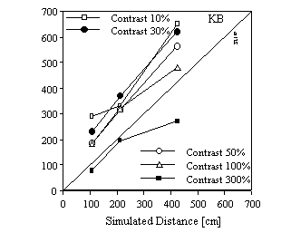 b
b