Perceived Quality of Video Enhanced for the Visually Impaired
Eli Peli
Vision Rehabilitation Service New England Eye Center, Tufts Medical School
and Schepens Eye Research Institute, Harvard Medical School
20 Staniford St. Boston, MA 02114
Tel: (617) 912-2597, Fax: (617) 912-0111
email: eli@vision.eri.harvard.edu
INTRODUCTION
Enhancement of video images was proposed as an aid for the visually impaired, and was shown to be effective using the Adaptive Enhancement algorithm in optical simulations (Peli and Peli, 1984), and in computational simulations of static images (Peli, et al., 1991). The same adaptive enhancement was demonstrated to improve recognition of static, black and white images of faces for patients with central scotoma and for patients with cataracts (Peli, et al., 1991). In a pilot study, Peli et al. (1994) found that 95% of patients preferred enhanced video segments when they compared them to the unenhanced version of the same segment. There was also a statistically significant, but small, improvement in recognition of details in still scenes taken from a motion picture. The enhancement in that pilot study was individually tuned for each subject by turning the two knobs controlling the Detail and Contrast levels in a free search procedure. In a later study, using a fixed enhancement for all subjects, Fine et al. (1997) found that only 21% of the subjects had preference for the enhancement. In both studies the preference was evaluated by questions following the viewing of both the enhanced and unenhanced versions of the same video segment. The current study tracked, continuously, the perceived quality of motion video viewed with and without enhancement. Both individual enhancement and arbitrary modification of the enhancement parameters were tested and compared with no enhancement as well as image degradation conditions. Patients selected the individual enhancement parameters in a more orderly procedure than in the pilot study. Preference was measured by continuously recording the patients' rating of the image quality in response to modifications of enhancement of a continuous video segment.
1. Individual tuning of enhancement
Twenty-three patients (ages 45-90, VA 20/100-20/800, 19 of them with documented central field loss) tuned the adaptive enhancement parameters by choosing a position on a bit-pad. The patients controlled the "Detail" parameter, representing spatial frequency, along one dimension of the pad and a combination of the "Contrast" and "Background" parameters on the other dimension. The axes and the direction of change varied in each trial. The DigiVision device (Hier, et al., 1993) was adapted to permit a no-enhancement condition and a reduction of contrast, resulting in low- pass filtering of the images.
The patients were presented blocks of 5 different digitally frozen video frames representing typical images encountered in movies. The order of the images within the block was randomized. Each block of images was presented a total of 4 times. The patient moved the mouse over the entire surface of the bit-pad while noting the changes in the image on the screen (a 27" Sony Trinitron TV monitor). The patient was asked to select the position which gave the optimal image based on two different criteria: a) "best " image overall (2 blocks), and b) "most details" visible in the image (2 blocks). Patients
viewed the screen with their habitual optical correction used for TV viewing at home. Sitting distance was adjusted to correspond to sitting distance at home (without use of binoculars) while accounting for the difference in TV set size as reported by the patient.
The data in Fig.1 represent the mean and SEM of the parameters selected by each patient averaged over 5 images, 2 criteria, and the 2 repetitions. The data show a clear correlation such that as the Detail setting (spatial frequency of the enhancement) decreased, the Contrast gain increased, but neither Contrast nor Detail settings were correlated with patient's visual acuity.
A 2(enhancement parameter) 5(image) ¥ 2 (criteria) 2(time) repeated measures ANOVA revealed a main effect of image [F = 4.117, p = .0044], a main effect of enhancement parameter [F = 22.470, p = .0001], and an interaction of image and enhancement parameter [F = 6.496,p = .0001] indicating a different setting for different images. Such interaction may be anticipated in consideration of the different spatial content which is relevant and interesting in different images.
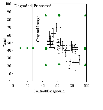
Figure 1 Mean enhancement parameters chosen by each patient (averaged across image, criteria, and time; i.e., 20 trials). Mean position correspond to mouse position on the bit pad. Error bars represent SEM. The vertical line at contrast = 27 (arbitrary units) represents no enhancement (original image). Area to the left of that line represents image degradation by low-pass filtering. Area to the right represents enhancement. All patients selected values corresponding to enhancement. The gray shaded points represent values used in the second part of the experiment for one subject indicated by the elipse.
Since there was no main effect of time or criteria, we decided that for the purpose of testing performance on a video detail recognition task, we would reduce the number of blocks to 2, each block consisting of only 4 images (excluding the night scene, for which responses varied the most). We repeated the same procedure using 2 blocks, 4 images, and only the "best" criterion, with another group of patients (n=29). One patient was unable to manipulate the bit pad due to arthritis. The results of 28 patients (Fig. 2) show a similar pattern. In this group also patients never selected no enhancement or low-pass filtered images. These findings indicate that we can measure preference for enhancement with static images, and that patients always preferred the enhancement over the un-enhanced images. Twenty of these patients took part in the second part of the experiment.
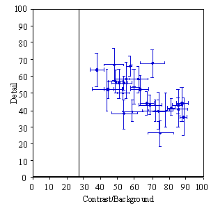
Figure 2 Enhancement parameters chosen by the second group of patients (averaged across image and time; i.e., 8 trials). Error bars represent SEM. The general pattern of result is the same as the results in Fig. 1. All patients preferred enhancement of some level and none selected no enhancement or low-pass version of the images. The same. correlation is apparent as found in the first group.
2. Perceived quality with motion video
To evaluate the perceived quality of color motion video we implemented a method similar to the method Hamberg and de Ridder (1995) used to evaluate dynamic changes in static imagery. This method enables continuous measurement of the perceived image quality as the display parameters (Details and Contrast) were changed. Ten elderly low vision patients participated (ages 48- 92 years, VA 20/100 - 20/500, all with central scotoma) Subjects indicated perceived quality with the adjectives: excellent, good, sufficient, poor, and bad by moving the bit-pad mouse on a scale printed in print large enough to be read easily. A beep every 10 sec. indicated a change in parameters and mouse position was recorded (once per sec.). A motion video segment played continuously (with no sound) while the enhancement parameters were abruptly switched between the following sets of 8 possible values:
•The selected set of parameters: Detail, D, and Contrast gain, C, chosen individually as described above (D, C), indicated in Fig. 1 for one subject by an ellipse around his data point.
• The original image (C = 1), indicated for the same subject with gray square on the vertical line.
• Intermediate values for both parameters (D, C/2); (D, C¥2); (D/2, C); (D¥2, C). The position of these settings are indicated by gray circles in Fig.1.
• Settings that degrade the image rather than enhance it by low pass filtering at 2 different levels of C<1 (contrast gains of 75% and 37%, respectively). The position of these settings are indicated by gray squares in Fig. 1. Each of these 8 settings was randomly presented within 10 blocks. The last 7 sec. per 10 sec. segment were averaged and the results of the 10 repeated presentations were averaged for each patient.
The results (Fig. 3) demonstrate that the subjects preferred the enhanced images over the un-enhanced image and preferred the un-enhanced image over the low-pass filtered images (DF=7, F=11.6, p<0.0001). Post hoc analysis showed that individual selection (D, C) resulted in statistically significant preference over the un-enhanced image (C=1) (DF=1, F=8.6, p=0.017). No differences were found between the individually selected set of parameters and the corresponding intermediate values.
A second group of 10 low vision patients was presented with other intermediate values.
• Intermediate values for both parameters (D/2, C/2); (D¥2, C¥2); (D/2, C¥2); (D¥2, C/2). The position of these settings are indicated by gray triangles in Fig.1. The second group was similar in composition (ages 65-89 years, VA 20/160-20/400, all with central scotoma) The second group had similar results, though the differences were smaller. For the combined group of 20 subjects, the main results remain significant and a significant correlation was found also between level of Contrast enhancement selected and patients' level of preference (r= 0.5, p= 0.023). The correlation with the Detail setting was not significant.
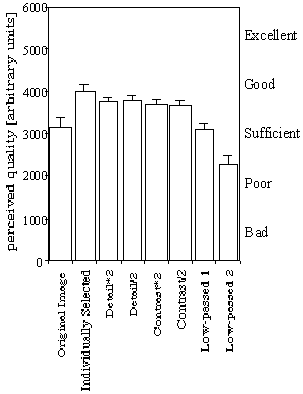
Figure 3 Perceived quality of motion video segments as indicated by 10 patients. As can be seen the image enhanced with the individually selected parameters (Fig. 2) provides the highest quality. The original un-enhanced image is perceived to be better than the low-pass filtered images. Various arbitrary modifications of the enhancement produced little difference in the perceived quality.
It is important to note that the patients as a group indicated a very small change in perceived quality of the images under all manipulation. While the quality of the original image was rated on the average as sufficient, the most degraded image which was quite severely degraded was rated less than one step below that (better than poor, on the average).
DISCUSSION
This study illustrates our ability to record a statistically significant effect in a continuous measure of preference for motion video. It further illustrates that the adaptive enhancement, (Peli and Peli, 1984) as provided by the DigiVision, adds substantially and significantly to perceived image quality. The fact that small group of low vision patients (n=10) is sufficient to demonstrate a statistically significant effect is an indication that the effect is robust. The variability in results across our studies may be accounted for by the methodological changes and possibly by variability in the groups population. The question of individual vs generic enhancement has not been answered definitively by this study. The variations between various levels and types of enhancement used in the study did not lead to statistically different effects. The individual selection did lead to a larger effect than all other conditions and the lack of significant difference may be due to the small number of subjects or the small level of variation used. It is clear, however, that patients with low vision can select a level of preferred enhancement which is consistent (small SD on repeat selection and across images) and follows a regular pattern for the group as a whole. We can also conclude that the enhancement selected using static images is preferred for the viewing of motion video. Patients who preferred stronger enhancement also perceived the enhancement to provide a larger benefit in image quality.
Paper Reference: Technical Digest on Vision Science and its Applications,
Technical Digest Series Vol. 1, 1999, in press (Optical Society of America,
Washington, DC).
ACKNOWLEDGMENTS
Supported by NIH grants EY05957, EY10285, EY10786. I thank Angela Labianca for her help on all aspect of the study. Charles Simmons programmed the bit pad control of the enhancement device. Rick Hier from DigiVision provided the specially modified system that could low-pass filter the images as well as enhance them.
REFERENCES
Fine, E., E. Peli, and N. Brady. Video enhancement improves performance of persons with moderate visual loss. In Proceedings of the International Conference on Low Vision, "Vision ‘96", Madrid, Spain, 85-92, 1997.
Hamberg, R. and H. de Ridder (1995) Continuous assessment of perceptual image quality. J. Opt. Soc. Am. A 12: 2573-2577.
Hier, R.G., G.W. Schmidt, R.S. Miller, and S.E. DeForest (1993) Real-time locally adaptive contrast enhancement: A practical key to overcoming display and human-visual-system limitations. SID 93 Digest 24: 491-494.
Peli, E., R.B. Goldstein, G.M. Young, C.L. Trempe, and S.M. Buzney (1991) Image enhancement for the visually impaired: Simulations and experimental results. Invest. Ophthalmol. Vis. Sci. 32: 2337-2350.
Peli, E., E. Lee, C.L. Trempe, and S. Buzney (1994) Image enhancement for the visually impaired: The effects of enhancement on face recognition. J. Optical Soc. of Am. A. 11: 1929-1939.
Peli, E. and T. Peli (1984) Image enhancement for the visually impaired. Optical Engineering 23: 47-51.