Testing a Simulation of Peripheral Vision
Using an Image Discrimination Task
Eli Peli
Schepens Eye Research Institute, Harvard Medical School, Boston, MA 02114
George A. Geri
Visual Research Laboratory, Raytheon Training Inc. Mesa, AZ 85212-0904
Abstract
The appearance of images in peripheral vision is an important consideration in the design of both wide-field and area-of-interest simulators, and can be simulated using vision models that account for the change in sensitivity as a function of retinal eccentricity. Simulations based on three different contrast sensitivity functions (CSFs), and a wide range of fundamental eccentricity constant (FECs) [cf., Peli et al., 1991, JOSA A, 8; 1762-1774] were tested here using an image discrimination task. Subjects were asked to discriminate an original image from a mirror image of the original processed at various fundamental eccentricity constant (FEC) levels. It was found that a combination of the foveal CSF and an appropriate FEC could characterize the discrimination of images with peripheral vision. The required FEC was found to be significantly larger for high sensitivity detection CSF as compared to the lower sensitivity orientation discrimination CSF. All FECs found here are larger than those computed from CSF measurements obtained across the retina. This difference may be due to masking by superimposed or adjacent image detail, which may result in a more rapid decline of detection performance as retinal eccentricity is increased (i.e., crowding or lateral masking effects).
Testing a Simulation of Peripheral Vision Using an Image Discrimination Task
Eli Peli
Schepens Eye Research Institute, Harvard Medical School, Boston, MA 02114
George A. Geri
Visual Research Laboratory, Raytheon Training Inc Mesa, AZ 85212-0904
Introduction
Simulating the appearance of a scene or image generated using a computational vision model can be a useful design and analysis tool. Validation of such simulations and underlying vision models are necessary. For instance, Peli (1996) tested and demonstrated the validity of a central (foveal) visual model (cf. Peli, 1990) using simulations of complex images. In that study, observers were asked to discriminate an original image from a simulation of the original as viewed from various distances. The distance at which discrimination performance was at threshold was compared with the simulated observation distance, and was found to be the same. Peli (1996) also determined that CSF data obtained with 1-octave wide Gabor patches in a detection task were the most appropriate to be used in the context of the tested vision model.
In the present study, we have used a more extensive visual model to simulate the appearance of various wide-field images, and we have performed a visual evaluation of those simulations. Previous work has suggested that the contrast sensitivity at various retinal eccentricities is dependent on both the foveal CSF and the fundamental eccentricity constant (FEC). The FEC is the rate of decrease in contrast sensitivity with retinal eccentricity as measured using a one c/deg grating (Peli et al., 1991). The purposes of the present study were to: 1) determine whether a single eccentricity-dependent parameter (i.e., the FEC) is sufficient to model the well-known spatial nonuniformity of the visual system, and 2) determine which of several CSFs is the best estimator of the discrimination of complex, wide-field imagery.
Methods
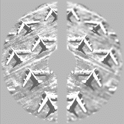
Stimuli and Apparatus. Four stimulus images were obtained from the left and right halves of two digitized aerial photographs, one of an airport and the other of planes on the ground. One half of each stimulus image was an unprocessed version of the original half-image, and the other was a mirror image of the original half-image (Fig. 1), processed as described below. Stimuli were rear-projected onto a large screen.
Simulations. To simulate their appearance across 64º of visual angle, the images were processed assuming fixation at their center. The details of the simulation method are given in Peli (1990), and the modifications used for peripheral simulations are given in Peli et al., (1991). The images were processed using one of three CSF data sets (gp, bc, or th) and one of seven FEC levels (0.02, 0.035, 0.055, 0.075, 0.10, 0.15, and 0.20). The FEC levels of 0.035 and 0.055 were found by Peli et al. 1991 to fit various peripheral CSF data sets from the literature. The remaining FEC levels were selected to cover a suitable range around these values. The gp CSF data were based on the discrimination of horizontal and vertical 1-octave Gabor patches (i.e., a sinusoid within a gaussian aperture) and were low-pass in character. The th CSF data were obtained using a contrast detection task and similar stimuli (Peli 1996). These were also low-pass in character. The bc CSF data were based on contrast detection of sinusoid gratings within a 2º square-wave aperture (Cannon, 1985). These data were band-pass in character and the absolute values for the mid spatial frequencies range was similar to that of the th data. The simulations where generated by assuming fixation at the center of the image. The image was decomposed into a multi-scale representation of local band limited contrast. Each pixel at each scale was tested against a threshold computed from the CSF the FEC and the pixel’s eccentricity.
|
|
Fig. 1. A typical stimulus. The image shown was obtained by applying an FEC level of 0.15 (the second highest) to the right side of the mirror-image of the right side of the "planes". The full stimulus images were 1024 x 1024 x 8-bits and subtended 64º at a viewing distance of 1.2 m. The central 12.8º of each image, a 3.7º vertical strip between the two half-images, and the surrounding area were replaced with a homogeneous field whose luminance was equal to the mean luminance of the stimulus images. The mean luminance of the stimulus images was about 1.2 fL.. The grayscale of each stimulus image was linearized (i.e., gamma corrected) using a look-up table. |
Testing Procedure. A 20-min. practice session was run for each of the observers on their first two visits. All sessions began with 8-10 min. of adaptation to a mean-luminance field. The stimulus image appeared 4 seconds after subject's previous response, and the observers were asked to indicate, by depressing either the right or left mouse button, whether the right or left half of the image appeared more blurred. The stimulus duration was 300 msec. The 560 trials run corresponded to 10 random presentations of each of 56 stimulus images (i.e., 4 original images x 2 sides for the standard x 7 FEC levels). Thus, for each session, forty responses were used to estimate a Percent Correct for each image type (airport or planes) at each of the seven FEC levels. Only one image set (either gp, bc, or th) was tested in each session.
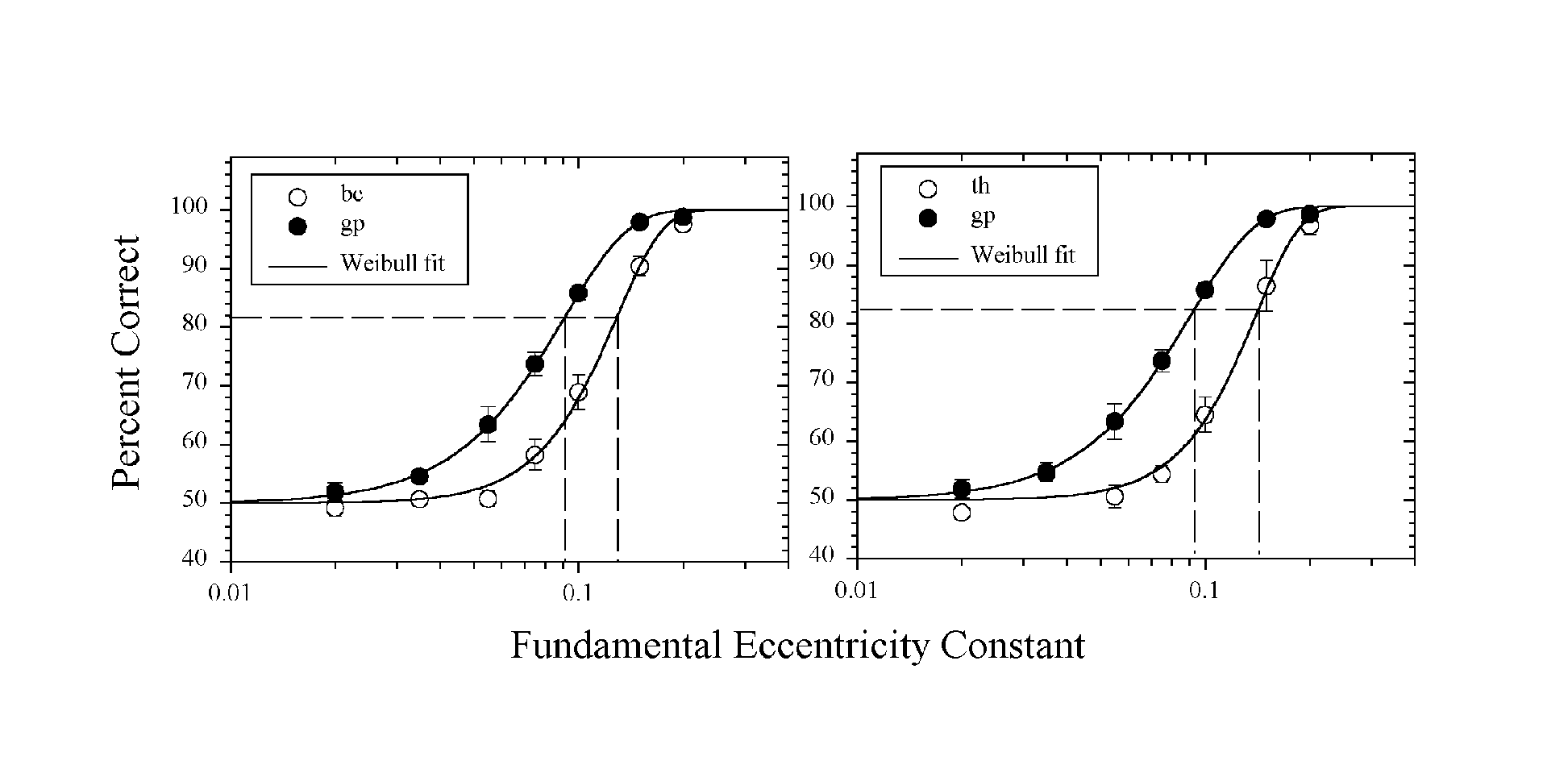
Data Analysis. The data presented here are means of five Percent-Correct estimates; each obtained from the forty responses within an individual session. The Percent-Correct data, plotted as a function of FEC level, were fitted with a Weibull distribution, which was used to estimate the FEC value corresponding to a Percent Correct of 81.6.
Results
Shown in the left graph of Fig. 2 are the functions relating Percent-Correct to FEC level for images simulated using either the bc (open symbols) or gp (closed symbols) CSF functions. The data represent averages for four observers. The smooth curves in the graph represent the best-fitting, two-parameter Weibull function. The FEC level corresponding to 81.6 Percent Correct was 0.128 for the bc data and 0.091 for the gp data. Analogous data comparing the results for the th and gp CSF functions are shown in the right graph of Fig. 2. The th data were obtained for three of the four observers from which the bc and gp data were obtained. The average threshold FEC level for the th data was estimated to be 0.140.
Discussion
Data analogous to those shown in Fig. 2 (left) have been reported by Peli and Geri (1993) and Peli (1995). In both of those reports, as well as in the present study, the FEC levels were chosen to span a level of about 0.50, which had been found by Peli et al. (1991) to fit various peripheral CSF data. The basic finding that the chosen FEC range resulted in a full psychometric function (see also Fig. 12 in Peli, 1995) indicated to us that the simulations were approximately correct. It would not have been surprising, with simulations based on only the foveal CSF and one eccentricity parameter, to find that the images were either all distinguishable or all indistinguishable from the original.
In the data presented by Peli and Geri (1993), one of the images resulted in a simulation that was easier to discriminate from the original. The model used for the simulations cannot account for this aspect of the data, which is observer-based and includes no image-related parameters. We have seen similar effects in testing simulations of central vision (Peli, 1996). In that case the effects were attributed to an artifact, the so-called high frequency residual (HFR), which was removed from the simulations but which remained in the original image. The HFR is the set of spatial frequencies at the corners of the square-shaped, spatial-frequency support, which are excluded when only a circularly symmetrical filter is used. Peli (1996) found that removal of the HFR resulted in the elimination of the image dependency as well as an improved performance of his simulations at various viewing distances. The data of Fig. 2 were obtained from images from which the HFR had been removed. With these stimuli, the data from only one observer showed a notable difference for the two images used (airport vs. planes). Further, the differences for this one observer tended to appear only for the more highly processed images, suggesting that the difference was due to a criterion difference in judging the images, rather than to differences in image detail attributable to the HFR.
|
|
|
Fig.2. A comparison of data obtained from images simulated using the bc and gp (left graph), or the th and gp (right graph) CSF functions. The smooth curves correspond to the best-fit, two-parameter Weibull function. The error bars are ±1 s.e.m. intervals about each data point. Note that in all cases at some FEC level the images are just distinguishable. The similarity between the th and bc results indicate that sensitivity to frequencies in the mid range of 1- 10 c/deg control the discrimination in this task. |
The data of Fig. 2 (left) show that wide-field images simulated using the bc and gp-based CSFs are discriminated at different FEC levels, thus extending the findings of Peli (1996) regarding the sensitivity of the simulation and testing method to small differences in CSF to peripheral vision. The most notable difference in the bc and gp CSFs is that the former are bandpass in character whereas the latter are lowpass. Since peripheral vision is relatively more sensitive to low spatial frequencies, it would be expected that images simulated based on the two types of CSF would be perceived differently—as was, in fact, found to be the case. However, as noted earlier, Peli (1996) found that simulations of foveal vision using Gabor patch stimuli better matched the appearance of the image from the simulated distance when the stimuli were based on threshold detection data as compared to orientation discrimination data. We therefore, added a third condition, th, resulting in a lowpass CSF but with a different absolute sensitivity. As can be seen from the data of Fig. 2 (right), the th data are similar to the bc data. Since the CSF of these two sets are very similar at intermediate spatial frequencies these results suggest that the differences in discrimination found in the present study are related to the absolute sensitivity of the underlying CSF function rather than to its shape. Further discrimination between the two functions may require testing with variable contrast images to probe lower spatial frequencies, as found in Peli (1999).
The vision model employed here produced images that were discriminable in a way predictable from well-established visual and perceptual data. It might, therefore, be expected that analogous models could be employed to assess more general image properties such as perceived image quality. In particular, the peripheral vision model suggested by the present analysis might be useful in evaluating wide field simulator images as well as area of interest, or other foveating systems.
Acknowledgments: This work was supported in part by grants EY05957 and EY10285 from the NIH, and by U.S. Air Force Contract F41624-95-C-5011 to Raytheon Training and Services Co. at the Air Force Research Laboratory, Mesa, Arizona. We thank Brian Sperry and Craig Vrana for programming support.
Paper Reference: Peli E. and Geri G. Testing the simulation
of peripheral vision with image discrimination. Technical Digest of Papers, SID-99, Digest,
1999; 424 - 427.
References
M.W. Cannon Jr. (1985) Perceived contrast in the fovea and periphery. J Opt Soc Am. A 2, 1760-1768.
E. Peli (1990) Contrast in complex images, J Opt Soc Am A 7, 2030-2040.
E. Peli, J. Yang, and R. Goldstein (1991) Image invariance with changes in size: The role of peripheral contrast thresholds, J Opt Soc Am A 8, 1762-1774.
E. Peli, editor, Visual Models for Target Detection and Recognition, (World Scientific Publishers, Singapore, 1995).
E. Peli, L. Arend, G. Young, and R. Goldstein (1993) Contrast sensitivity to patch stimuli: Effects of spatial bandwidth and temporal presentation, Spatial Vis 7, 1-14
E. Peli (1996) Test of a model of foveal vision by using simulations. J Opt Soc Am A 13: 1131-1138
E. Peli (1999) The contrast sensitivity function (CSF) and image discrimination. SPIE Proceedings Human Vision and Electronic. In press.
E. Peli and G. Geri (1993) Putting simulations of peripheral vision to the test. Invest. Ophthalmol. Vis. Sci., 34 (ARVO Suppl.), 820.