
TREATING HEMIANOPIA USING PRISMS TO CREATE PERIPHERAL DIPLOPIA
Eli Peli
The Schepens Eye Research Institute, Harvard Medical School
20 Staniford Street, Boston, MA 02114
Abstract
Most optical aids for hemianopia provide only field relocation while field expansion is the desired effect and thus, these aids are rarely successful. This paper describes a novel method for prism correction of hemianopia that provides actual field expansion in a convenient and functional format. To expand the upper quadrant of the field, a high power prism segment (30-40 PD) is placed base-out across the upper part of the spectacle lens, on the side of the loss, at about the level of the limbus. A similar prism segment at the lower part of the lens is used for the lower field. Usually the upper prism is applied first, as adaptation is easier. The patient is instructed to foveate through the carrier lens only, not through the prisms, and to use vertical head movements to explore the lower field foveally. The peripheral location of the prisms causes peripheral diplopia as wide as the field of the prism and shifted by 15-20 deg. relative to the field of the other eye. At the edge of the hemianopic field loss, objects that would fall in the scotoma of one eye are seen through the prism in the other eye, providing a real field expansion. The field expansion is provided at any position of gaze, including gaze away from the side of the scotoma. The effect of the prism on field expansion has been demonstrated using binocular Goldmann perimetry. Most patients reported improvement in function and obstacle avoidance. Patients with hemianopia and abnormal binocular function (strabismus or amblyopia) reported no noticeable effect of the prism during the trial period, suggesting that the method’s effect is limited to patients with normal binocular vision. An approach for fitting the system to patients with abnormal binocular vision was developed as well.
Introduction
Homonymous hemianopia, the loss of half the visual field on one side in both eyes is a frequent consequence of brain damage from stroke, head injury, or surgery to remove tumors. Hemianopic field loss causes problems in mobility and navigation. Patients frequently complain of bumping into obstacles on the side of the field loss and getting bruised on their arms and legs. The number of such accidents may decrease with adaptation to the condition, presumably because patients become more cautious and learn to use head and eye scanning techniques to avoid the collisions. Despite such improvements, many patients continue to suffer from the effects of limited visual field .
Various devices have been considered and applied for the management of hemianopic field defects including mirrors, partially reflecting mirrors (beam splitters), prisms, and reversed telescopes . The effects of these devices may be classified as providing either field relocation (shifting) or field expansion. Field expansion is the desired effect, as it means that the simultaneously seen field is larger with the device than without it. Field relocation only exchanges the position of the field loss relative to the environment or relative to the body’s midline. This means that a part of the environment that was obscured by the scotoma becomes visible, but at the same time a different part of the environment (of the same angular span) which was visible becomes invisible. This may be useful under some circumstances and assumptions, but clearly it is inferior to field expansion.
Binocular sector prisms , the most commonly used technique (Fig.1), provide only for field relocation. In this case the prism (in the form of a Fresnel press-on, glued segment, or molded lens) is limited to the part of the carrier lenses which would be in the scotomatous field when the eyes are in primary gaze position. Their effect, therefore, is limited to instances when the line of sight is directed through the prism sector of the spectacle lenses. When the patient is looking through the other part of the lens the prism has no effect, as it is included completely in the scotoma. When looking through the prism sector the field is shifted. Since the patient does not see objects in this part of his field, he is less likely to fixate into this field and intentional scanning is required. Furthermore, the optical field loss caused by a binocular sector prism (Jack-in-the-box scotoma) ) is in the center of the field of view. This field loss can be compensated for by head movements and in part by eye movements, but is a serious deficit in the design of these aids. The amount of prism typically used with this technique provides a very small shift of the field (6 - 9 deg.). Comparable access to the unseen part of the field could be achieved with a slightly larger eye movement than is necessary to shift into the field of the prism.
Monocularly fitted sector prisms expand the field once the patient changes his fixation to within the field of the prism. As long as the patient's eyes are at primary position of gaze or are directed away from the hemianopic field side, the monocular sector prism has no effect on the field of view. Diplopia and confusion accompany the field expansion, as a result of directing the gaze into the field of the prism. Confusion refers to the appearance of two different objects at the same perceived direction. Confusion in this case represents the intended beneficial effect, as it represents the appearance of an object that would be invisible without the prism. However, the central diplopia accompanying the confusion is very unpleasant to the patient and may account for the lack of success.
Successful design of such a hemianopic visual aid requires that it expands the field rather than relocates it, functions in all positions of gaze, and avoids central diplopia. These considerations led me to develop a new method of field expansion described here. The new method involves a monocular sector prism that is limited to the peripheral field (superior, inferior, or both). It is placed across the whole width of the lens spanning both sides of the pupil (Fig. 2) so that it is effective at all lateral positions of gaze. The prism expands the field via peripheral diplopia and confusion. Peripheral diplopia, however, is much more comfortable for the user than central diplopia since peripheral physiologic diplopia is a common feature of normal vision . The field expansion effect of the prism is unaltered by lateral eye and head movements over a wide range of such movements into either side.
In fact, the prisms used by this method render the patients "exotropic" in peripheral vision while leaving the foveal vision unaffected. The constant exotropia provides the field expansion similar to that enjoyed by exotropic congenital hemianopes . Through the adaptation of peripheral vision and the change in perceived direction reported by Kohler it enables the field expansion to be useful and functional. Furthermore, the chromatic aberrations of the prisms provide a spectral cue that distinguishes the objects viewed through the prism from those seen through the clear carrier lens. This spectral difference facilitates adaptation to the prism by reducing the ambiguity associated with the confusion, clearly marking the objects as to their eye of origin.
The peripheral prism design described here provides for a field expansion that is measurable by standard perimetry. In addition, since the prism only affects peripheral vision, a higher power prism can be used despite its inferior optical quality. Fresnel prisms of 40 prism diopters have been used. The field expansion of about 20 deg. provided by these prisms at the middle of the field, in all positions of gaze, was found to be very effective in providing the patients with the necessary view needed to avoid obstacles while walking. Also, patients reported an adaptation leading to veridical perception of the direction of objects detected with the prisms; however, no formal testing of the adaptation has been conducted.
Fig.1. Binocular sector prisms as are commonly prescribed for right homonymous hemianopia. The prisms with base to the right are placed on both lenses' right sides up to the pupil. Note that these prisms have no effect when the patient eyes are at primary position of gaze or are looking left (where all visual stimuli come from). When the patient looks through the prism the field is just SHIFTED not enlarged. Furthermore, these prisms cause an optical field loss at the center of the lens.
Methods
Patients
Twelve patients were evaluated. Six had homonymous hemianopia following strokes and surgeries to remove lesions (5 left hemianopia). Two had hemianopia secondary to surgery to remove lesions (1 left hemianopia). One had left hemianopia due to head trauma. Three had homonymous quadrantanopia. One of theses was only partial and limited to the central part of the lower left quadrant (Fig. 4A) following surgery to remove malignancy. One patient had a lower right quadrantanopia secondary to seizures and another had upper quadrantanopia following surgery to stop seizures. Ages ranged from 41 to 75 years. Visual acuity was good in both eyes of all patients, varying from 20/15 to 20/30.
Prism placement and mode of operation
Separate prism segments are used to expand the upper and lower quadrants. To expand the upper quadrant of the field the base-out prism segment is placed at the upper part of the spectacle. The prism segment is placed across the upper center of the spectacle lens with its lower edge above the pupil at about the level of the limbus and covers as much of its width as is practical (Fig. 2). Similarly, a prism segment at the lower part of the lens is used for expansion of the lower field.
Most patients were fitted with Fresnel press-on prism segments of 40 D (3M Press-on prisms). These soft plastic segments are cut from a larger piece and fitted to the frame shape to cover the full width of the lens. Two patients were fitted with a permanent elliptical prism segment inserted inside the spectacle carrier lens (Fig. 2 upper segment). While this type of segment has better optical qualities than the Fresnel segment, it has a number of disadvantages. The cost is substantially higher, the thick prism base protrudes dangerously close to the eye, and the size of the segment is therefore limited and cannot cover the full width of the carrier lens. For these reasons and since the optical quality is not critical in peripheral vision, where the prism is used, I have discontinued using these segments in more recent cases.
Since the prism is placed across the upper center of the carrier lens, it effects the patient in all positions of gaze. The patient is instructed to foveate through the carrier lens only and not through the prism. Looking through the prism causes central diplopia that is bothersome and uncomfortable. For a patient with normal vision (no hemianopia) the peripheral location of the prism would provide a peripherally diplopic field as wide as the field of the prism and shifted by 15-20 deg. relative to the field of the other eye. For patients with hemianopia, the situation is similar over a large portion of this field (corresponding to the non-scotomatous field); objects are seen in diplopia providing no particular help. At the edge of the field, however, objects that fall in the scotoma of the eye without the prism are seen through the prism in the other eye providing a real field expansion of about 15-20 deg. This additional field is provided for any position of gaze including gaze away from the side of the scotoma. Rather than diplopia, the objects that come into view due to the prism are seen in confusion since two objects (seen by the two eyes) are perceived to be in the same direction. Confusion always co-exists with diplopia and therefore it is also part of physiological diplopia. Thus, we are as accustomed to confusion in our peripheral fields as we are to diplopia.

Fig. 2. An illustration of the new prism correction for left hemianopia. The prisms are worn only over the left eye. They are restricted to the upper and lower peripheral fields but extend across the whole lens so that they are effective at any position of lateral gaze. Illustrated are a simple mounted prism for the upper segment and a Fresnel press-on prism on the lower segment.
Visual field testing
Most previous methods using prisms to treat hemianopia result in field relocation rather than field expansion. Methods such as the binocular full prisms and the binocular sector prisms do not result in an expansion of the visual field as measured by perimetry. In fact binocular sector prisms cause an optically induced scotoma at the center of the lens. Monocular sector prisms do expand the visual field when the gaze is directed into the field of the prism but usually this field does not include the primary position of gaze as commonly used in perimetry. As a result most previous prism based treatment methods did not attempt to demonstrate field expansion by standard perimetry. Many investigators claimed that the prism provided an increase in field "awareness" which could not be measured by perimetry, but was noted by the patients.
The peripheral prism design described here provides for a field expansion, which should be measurable by standard perimetry. In addition the magnitude of the expansion can be predicted based on the prism power, and can be tested by the perimetry. The field expansion should occur at all positions of gaze and therefore it should be measurable at the primary position of gaze.
We first measured patients’ fields monocularly using standard perimetry techniques to obtain a description of their field loss. Following prism prescription, the patients’ fields were measured binocularly with and without the prism using a standard Goldmann screening. The Goldmann fields were measured using a standard screening procedure consisting of dynamic mapping and static perimetric probing within identified non-scotomatous areas. However, the patient viewed the fixation target binocularly and the chin rest was centered to keep the head in the center of the bowl. Fixation was monitored through the observation tube in the common way.
Results
Field expansion
For all patients (but one, discussed below) the field measured binocularly with the prism segment demonstrated the expected field expansion in the corresponding quadrants (about 20-30 deg.) (Figs. 3 and 4). This field expansion could also be demonstrated in confrontation fields where it also demonstrated to the patients (and their present family members) the extent of the field expansion and the effects on obstacle detection in free space.
Subjective effect
The first two patients have been followed for more than a year now. Both (one with hemianopia and one with upper quadrantanopia) reported pleasure with the beneficial effect of the prism. Following a short adaptation period of 2 weeks to the upper segment and 3 weeks for the lower segment, they could use it as instructed and noticed that it expanded their field and prevented accidents. These two patients were fitted with permanent prescription incorporating the elliptical prism segment. After more than one year they remain very pleased with the correction. Both patients are driving. One is driving following a Department of Motor Vehicle evaluation. The upper quadrantanopia patient has been approved for driving since his horizontal field of view was sufficient and he has been free of seizures for more than 6 months. He still feels safer and more comfortable driving with the prism segment.
One patient with right lower quadrantanopia has been followed for more than a year. This patient is emmetropic and needs to wear plano spectacles to be able to use the prism. This patient is very pleased with the effect and continues to wear the plano correction on a full time basis. He noticed a substantial reduction in his rate of bumping into obstacles and requested to have a spare spectacle fitted with the correction. Except for the 2 patients reported below, all of the other patients are very pleased with the effects of the correction. They reported rapid adaptation to the prisms within two to three weeks and noted significant improvement in obstacle avoidance. Two patients have stopped using the (short) canes they were using before the treatment as their confidence increased with the prisms.
One of these patients, whose fields are illustrated in Fig. 4, helps demonstrating the importance of the paracentral 20 degrees (provided by these prisms) for safe mobility. This patient’s visual loss was a homonymous partial lower left quadrantanopia (Fig. 4A). Despite a fairly complete peripheral field, this patient reported substantial difficulties with bumping into obstacles, especially furniture. This is reasonable to expect as obstacles for mobility are likely to be fairly close to the midline and to the line of heading of a walking person, or they will not intersect with the path. Realizing that this is the case also explains why the prisms provided by us, which expand the field only by 15- 20 degrees, are found by the patients to be so effective.
Two patients with hemianopia had no subjective complaints before treatment. They knew of their field loss from their doctors’ reports and could demonstrate it for themselves, but had adapted to the situation to such an extent that they had noticed no limitation on function. These two patients were fitted with prism segments for a trial period. In both cases the field expansion with the prism was demonstrated with perimetry as expected. Both patients, however, reported no benefit from the prisms but also had no particular difficulty due to the prism. Since no positive effect was achieved the prism wear was discontinued.
Patients with abnormal binocular vision
A
B
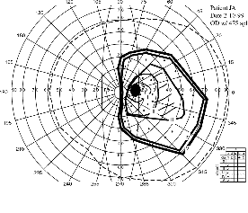
Fig. 3. The visual field of a patient with left hemianopia (A) measured in the right eye (left eye similar), and (B) measured binocularly with the prism correction (40 PD) in both upper and lower segments before the left eye. Note the additional field extending about 20 degrees left of the midline with the prism correction.
One patient with hemianopia, who had severe complaints, after an initial trial period with prisms reported no subjective improvement with the prisms nor any difficulties wearing them. To my surprise, this patient demonstrated little change of the binocular field measured when wearing the prisms. On further examination, it was determined that this patient had a third nerve palsy as another consequence of her stroke. She had constant strabismus and was suppressing the eye on the side of the scotoma. The lack of functional binocular vision explains the lack of effect of the prism designed to work under binocular conditions. A second patient with left hemianopia had a history of amblyopia in the right eye, no strabismus and only weak intermittent indication of central suppression. During the trial period, she reported that the peripheral view through the prism segment on the left lens was dominant and resulted in erroneous perception of the direction of objects in the upper field. The effect was bothersome when traveling as a passenger in a car and when walking in the forest. This patient decided not to continue the trial. The results with these two patients seem to indicate that the method may not be effective for patients with abnormal binocular function. A third patient with severe diplopia raised a possible solution for this limitation. This patient had left hemianopia due to head trauma as a result of a bicycling accident. The same injury also caused left orbital trauma and scarring resulting in constant diplopia. To resolve the diplopia, this patient frosted the left lens with nail polish. I removed the nail polish from the peripheral portions of the lens where the prism segments were fitted and left it at the center. This arrangement provided for the peripheral field expansion as in the other cases but avoided the central diplopia caused by the orbital injury. This system worked satisfactorily for the patent and could be useful in any case of binocular disfunction. In fact in cases of exotropia together with hemianopia, which are quite common, the treatment may include only the blocking of the central portion of the lens with a translucent tape or nail polish coating. This will provide peripheral field expansion and will resolve the central diplopia at the same time. Note that the patient in such a case will continue to have central vision from the fixating eye and will only lose the central vision in the deviating eye.
A
B
(click on figure to zoom)
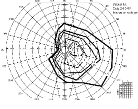
Fig. 4. Visual fields of a patient with partial left lower quadrantanopia secondary to surgery to remove a brain tumor. (A) The visual field of the right eye was similar to that of the left eye (not shown). Note the absolute scotoma near fixation. The fact that this patient had serious difficulties with avoiding obstacles, despite an almost complete field, emphasizes the importance of the near central field. The prisms, by compensating for the scotoma, provided safe mobility. (B) The visual field measured binocularly with the prism correction (40 PD), only in the lower segment, in this case resulted in complete elimination of the scotoma and relief of symptoms.
Discussion
This novel method for prism device for hemianopia provides actual field expansion in a convenient and functional format. Fresnel press-on prisms were useful for both the evaluation of the effect and for constant wear. Using the approach in both the upper and lower peripheral fields is effective and provides expansion of much of the peripheral field over a wide field of gaze. The method has been tested with patients with varying conditions and etiologies. It provides relief of symptoms to patients with normal binocular vision who have difficulties due to the hemianopia. Patients with strabismus (especially exotropia) may benefit from blocking of central vision in the eye on the side of the field loss.
Future work requires formal evaluation of treated patients in mobility situations to verify the reported effect of increased obstacle avoidance. Proper use of the prism in mobility requires adaptation that will correct for the distorted apparent directions of objects caused by the prism. The existence of such adaptation should be determined and after- effects evaluated. With the prism correction hemianopes may be able to drive more safely. The effects of this correction on driving performance needs evaluation as well, preferably in driving simulators. Because the prisms are used in peripheral vision, even stronger prisms may be useable than the currently available 40 D. Such prisms need to be evaluated.
Acknowledgements
Supported in part by NIH grants EY10285 and EY09597. I thank Dr. T Hedges for referring his patients to me and encouraging me through the development and testing of this method.
References
Bishop, P. O. (1981). Binocular vision. In R. A. Moses (Ed.), Adler's Physiology of the Eye: Clinical Applications, C.V. Mosby, St. Louis. 575-649.
Cohen, J. M. and Waiss, B. (1996). Visual field remediation. In R. G. Cole and B. P. Rosenthal (Eds.), Remediation and Management of Low Vision, Mosby, St. Louis. 1-25.
Faye, E. D. (1976). Characteristics of near vision aids: Convex lenses, telescopic loupes, closed-circuit television., Clinical Low Vision, Little, Brown and Co., Boston. first edition edn, Vol. 8, 63-85.
Goodlaw, E. (1983). Review of low vision management of visual field defects. Optometric Monthly 74, 363-368.
Göte, H., Gregersen, E. and Rindziunski, E. (1993). Exotropia and panoramic vision compensating for an occult congenital homonymous hemianopia: A case report. Binocular Vision & Eye Muscle Surgery Qtrly 8, 129-132.
Gottlieb, D. D. (1988). Method of using a prism in lens for the treatment of visual field loss, US. Patent No 4, 779, 972.
Gottlieb, D. D., Allen, C. H., Eikenberry, J., Ingall-Woodruff, S. and Johnson, M. (1996). Living with Vision Loss, St. Barthelemy Press, Ltd., Atlanta, GA.
Herzau, V., Bleher, I. and Joos-Kratsch, E. (1988). Infantile exotropia with homonymous hemianopia: A rare contraindication for strabismus surgery. Graefes Arch Clin Exp Ophthalmol 226, 148-149.
Jose, R. T. and Smith, A. J. (1976). Increasing peripheral field awareness with Fresnel prisms. Optical Journal and Review of Optometry 113, 33-37.
Kohler, I. (1964). The formation and transformation of the perceptual world. Psychol Issues 3(4), 14-173.
Rossi, P. W., Kheyfets, S. and Reding, M. J. (1990). Fresnel prisms improve visual perception in stroke patients with homonymous hemianopia or unilateral visual neglect. Neurology 40, 1597-1599.