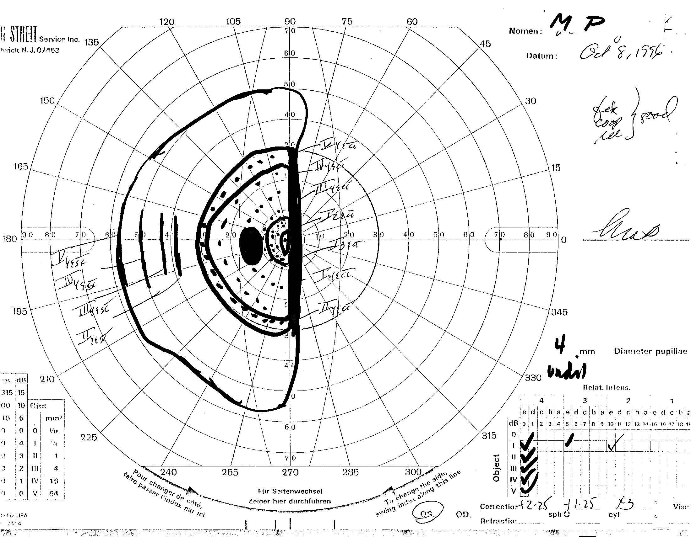
a (click on image to zoom)
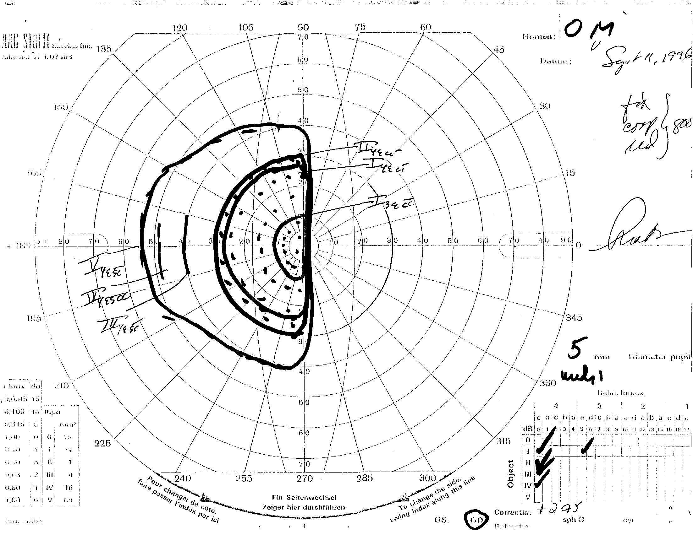
a (click on image to zoom)
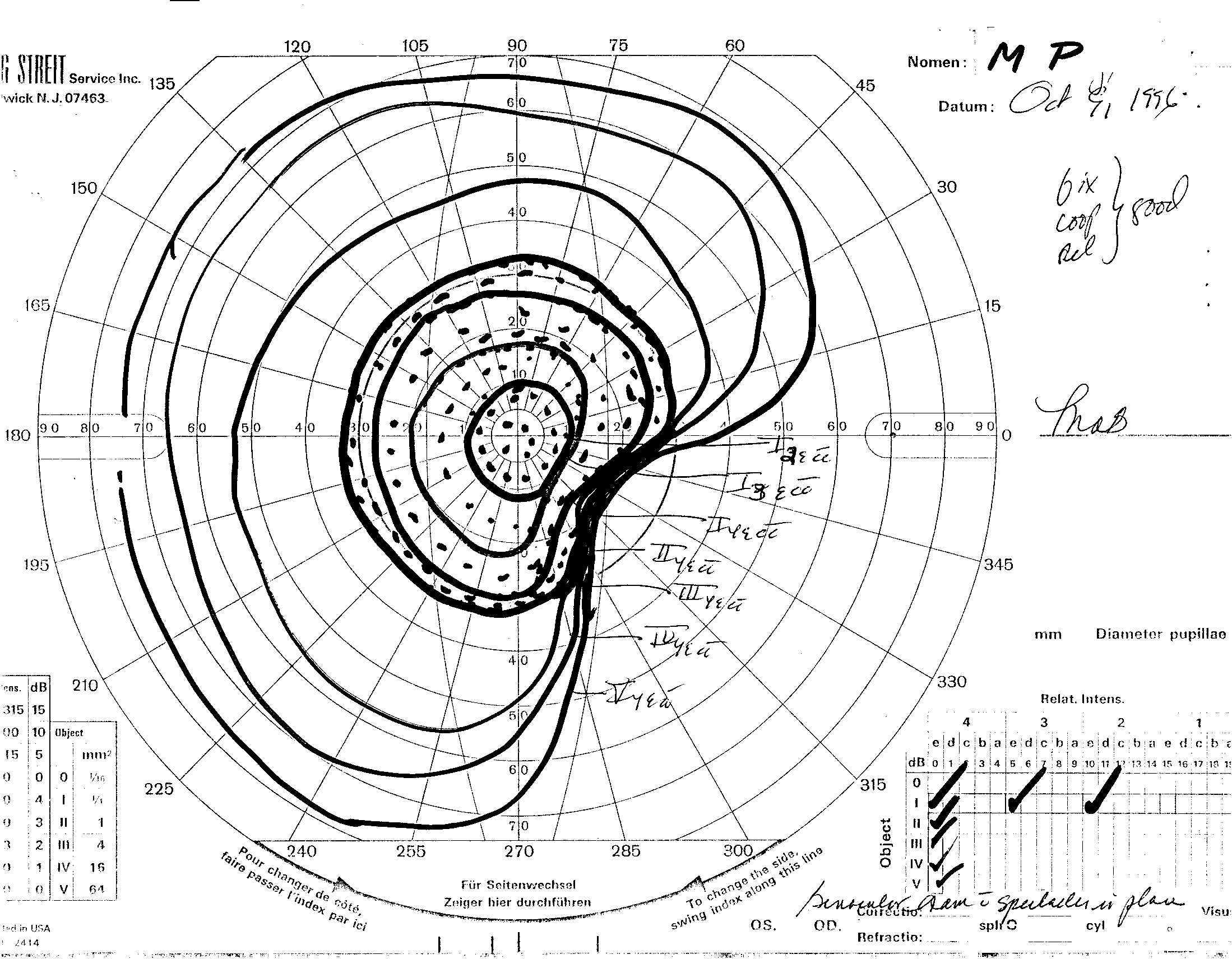
b (click on image to zoom)
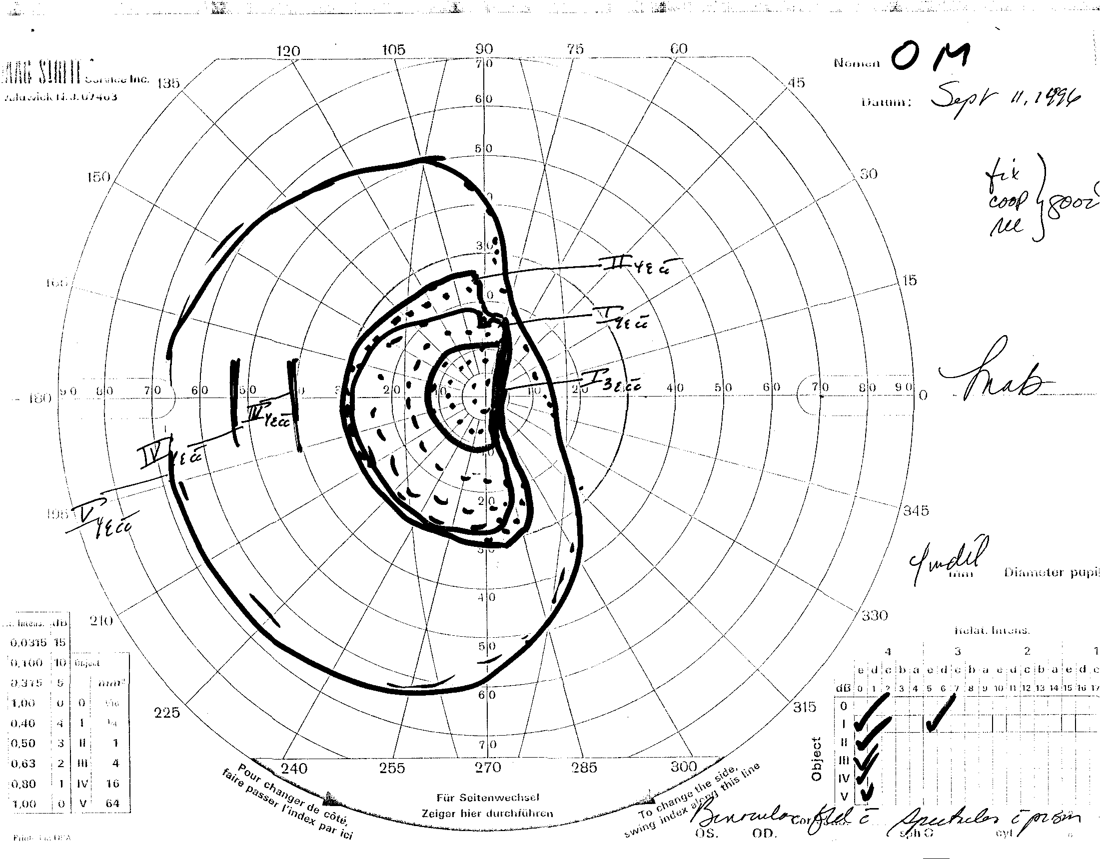
b (click on image to zoom)
Field Expansion for Homonymous Hemianopia using Prism and Peripheral Diplopia
Eli Peli
The Schepens Eye Research Institute, Harvard Medical School
20 Staniford Street, Boston, MA 02114
Tel: 723-6078 x597 Fax: 523-3463
Introduction
Visual field loss following a cortical lesion from stroke, brain surgery, or head trauma frequently manifests as homonymous hemianopia. Hemianopic field loss causes problems in mobility and navigation as well as reading. Patients frequently complain of bumping into obstacles on the side of the field loss and getting bruised on their arms and legs. The number of such accidents decreases with adaptation to the condition, presumably because patient are getting more cautious and use head and eye scanning techniques to avoid the pain. Despite such improvements many patients continue to suffer from the effects of limited visual field. Of great concern to many patients and vision rehabilitation personnel is the question of driving with such field defect. In all states where visual field requirements for driving are legislated, such field loss will disqualify drivers, unless qualified by special testing and licensing.
Clinical techniques which have been considered for the management of peripheral field defects include utilization of devices such as mirrors, partially reflecting mirrors (beam spliters), reversed telescopes and prisms (Goodlaw, 1983). In addition, training in scanning and awareness enhancement (Cohen & Waiss, 1996) is frequently attempted.
The effects of various devices can be classified as field relocation or field expansion. Field expansion is actually the desired effect as it means that the simultaneously seen field is larger with the device than without it. Field relocation only exchanges the position of the field loss relative to the environment. This may be useful under some circumstances and assumptions, but clearly is inferior to field expansion.
Field relocation occurs with the use of binocular opaque sector mirrors or with the use of overall binocular prism (Cohen & Waiss, 1996). Field relocation with overall binocular prism assumes that the eyes do not compensate for the prism (usually about 20 prism diopters) with a corresponding eye movement of about 10 degrees which will neutralize the effect of the prism. The effect of the prism on observers with normal vision, however, suggests that such compensation, and even adaptation, does take place. There is also a corresponding field loss in the far periphery on the seeing side occurring even if eye movements negate the beneficial effect of the prism. The field loss caused by a binocular sector prism (Jack-in-the-Box scotoma) (Cohen & Waiss, 1996) is in the center of the field. This field loss can be compensated by head movements and partially by eye movements. This approach also provides only for field relocation. Furthermore, the effect of the prism takes place only after the patient changes his fixation towards the side of the hemianopic field. Since the patient does not see objects in this part of his field, he is less likely to fixate into this field and intentional scanning is required. The amount of prism typically used provides a very small shift of the field. A comparable access to the unseen part of the field could be achieved with a slightly larger eye movement than is necessary to shift into the field of the prism. Binocular sector mirrors do provide view of a segment in the hemianopic field which can be large and positioned quite far into the periphery, but it is traded with an equal size scotoma on the seeing side.
Monocularly placed sector mirrors provide an actual field expansion, since the part of the hemianopic field seen in the mirror in one eye is superimposed on the seeing part of the field of the other eye. Of course that means confusion as two different objects are perceived to be in the same direction. The image reflected in a mirror is inverted right to left and is projected into the opposite direction of where it really is, making use of the device very difficult. Similar effects, with similar difficulties, can be achieved over a larger field (and for monocular patients) using semi-reflective mirrors. Here, in addition to the previously listed problems, the reflected image is lower in contrast. Monocularly fitted sector prisms expand the field, once the patient changed his fixation to within the field of the prism. As long as the patient's eyes are at primary position of gaze or are directed away from the hemianopic field the monocular sector prism (Jose & Smith, 1976; Gottlieb, 1988) has no effect on the field of view. The field expansion, achieved upon directing the gaze into the prism in the direction of the hemianopia, is accompanied by diplopia. The central diplopia induced is very unpleasant to the patient and may account for the lack of success (Cohen & Waiss, 1996). As can be seen, much of the difficulties with various field remidiation schemes is a result of insufficient consideration of the dynamic nature of the situation. The effects of eye and head movements need to be considered in the design and use of such devices.
These limitations and the lack of success with these devices in clinical practice has led me to look for a new method of field expansion. The new method involves a monocular sector prism which is limited to the peripheral field (i.e., superior, inferior or both). The peripheral prism is placed across the whole width of the lens spanning both sides of the pupil so that it is effective at all lateral positions of gaze. The prism expands the field via peripheral diplopia. Peripheral diplopia, however, is much more comfortable for the user than central diplopia since peripheral diplopia is a common feature of normal vision. The field expansion effect of the prism is unaltered by eye and head movements over a wide range of such movements into either side.
Methods
Separate prism segments are used to expand the upper and lower quadrants. To expand the upper quadrant of the field at all positions of gaze, a prism segment is placed base-out at the upper part of the spectacle lens on the side of the loss. The high power (30-40
D) prism segment is placed across the center of the spectacle lens above the pupil at about the level of the limbus and covers as much of its width as is practical (Fig. 1). Similarly, a prism segment at the lower part of the lens may be used for expansion of the lower field. We have not implemented the lower prism with patients yet, since it appears to be a much more complicated position to address. We decided to gain more experience first with the upper correction.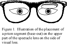
The prism is placed across the center of the lens, and thus affects the patients in all positions of gaze. The patient is instructed to foveate through the carrier lens only and not through the prism. Looking through the prism causes central diplopia which is bothersome and uncomfortable. The peripheral location of the prism should provide a peripherally diplopic field as wide as the field of the prism and shifted by 15-20 deg relative to the field of the other eye. Over a large portion of this field (corresponding to the non scotomatous field) objects are seen in diplopia providing no particular help. At the edge of that field, however, objects that would fall in the scotoma of one eye are seen through the prism in the other eye providing a real field expansion of 15-20 deg over the height of the prism segment. This additional field is provided for any position of gaze including gaze away from the side of the scotoma.
The patient is instructed that when an object of interest is detected through the prism (in peripheral vision) it should be examined through the carrier lens which requires head movement. In normal vision such head movements are preceded by foveating eye movement. The patient's training consists of learning to avoid the eye movement or to follow them quickly with head movement to eliminate the diplopia if it occurs.
For the initial trial period patients were fitted with a Fresnel press-on prism segment. The 40
D base-out segment is cut to fit the top part of the spectacle lens on the side of the visual field loss. The segment is trimmed to the height of the limbus margin. The patient is instructed in the care of the prism and in the use of the prism. On a follow up visit, following a trial period of 2-3 weeks, the patients were interviewed about the effect of the prism and their experience with it. The patient's binocular visual field with the prism segment was tested as well. Successful patients were fitted with a permanent elliptical prism segment mounted inside the spectacle carrier lens. Such segments can be ordered from Multilens Optical Solutions in Mölnlycke, Sweden.Four patients were evaluated with the Fresnel prism segments. Three had homonymous hemianopia following strokes and one had homonymous upper quadrantanopia secondary to brain surgery to treat seizures. All were particularly interested in the field expansion in relation to driving and all reported occasionally bumping into obstacles on the side of the scotoma.
Results
The effect of the prism on field expansion has been demonstrated using binocular Goldman perimetry. The patient's field at each eye was recorded using a standard Goldman screening technique consisting of dynamic mapping and static perimetric probing within identified non scotomatous areas. Examples of the monocular fields recorded are shown in Figures 2a and 3a. A binocular field was recorded with the patient wearing the spectacles including the Fresnel prism segment.
For 3 of the 4 patients the field measured with the prism segment demonstrated the expected field expansion in the upper quadrant of about 20-30 deg (Fig. 2b). To our surprise one patient demonstrated little change of field with the use of the prism (Fig. 3b). This same patient also reported no noticeable effect of the prism during the trial period. On further examination, it was determine that this patient had a third nerve palsy as another consequence of her stroke. She had constant strabismus and was suppressing the eye on the side of the scotoma. The lack of functional binocular vision explains the lack of effect of the prism designed to work under binocular conditions.
Two patients (one with hemianopia and one with the upper quadrantanopia) reported pleasure with the effect of the prism. Following an adaptation period they could use it as instructed and notice that it does expand their field and prevented accidents. These two patients were fitted with permanent prescription incorporating the elliptical prism segment. The patients with permanent correction have been followed for almost one year and are still very pleased with the correction. Both patients are driving. One is driving following a DMV evaluation. The quadranopia patient has been approved for driving since his horizontal field of view is intact and he has been free of seizures for more than 6 months. He still feels safer and more comfortable driving with the prism segment.
The fourth patient had history of amblyopia in the right eye. She had no strabismus and only weak intermittent indication of central suppression. During the trial period, she reported that the peripheral view through the prism segment on the left lens was dominant and resulted in erroneous perception of the direction of objects in the upper field. The effect was bothersome when traveling as a passenger in a car and when walking in the woods. This patient decided not to continue the trial.
Conclusion
A novel method for prism correction of hemianopia provides actual field expansion in a convenient and functional format. It's use is limited for patients with intact binocular function. The use of Fresnel press-on prism is useful for the evaluation of the effect. A permanent prism inset can be prescribed for successful users. The development of a multi segment lens that can cover wide field of gaze with high power prisms needs further development. The possibility of using the same approach in the lower peripheral field needs to be tested. If effective, it can provide expansion of most of the peripheral field over a wide field of gaze, providing significant help for patient with homonymous Hemianopia.
Supported in part by NIH grants EY10285 and EY09597.
Paper Reference: Technical Digest on Vision Science and it Applications, Technical Digest Series Vol 1, 1998, 74-77 (Optical Society of America, Washington, DC).